Anatomy Of Chest And Ribs - Thorax - Surface Anatomy, 4 Edition - True, false and floating ribs are denoted.. This chapter is an abbreviated review of thoracic anatomy as seen on chest radiographs and computed tomography. In vertebrate anatomy, ribs (latin: Xiphoid surgery relieves mysterious chest pain for young patient. O bones—spine, ribs, clavicles, scapulae, humeri. Anatomy of the cardiac chambers.
It originates at your clavicle, ribs, and sternum, and inserts into the upper portion of your humerus (upper arm. They are twelve in number on either side; Spiral ct of thoracic inlet. In most tetrapods, ribs surround the chest, enabling the lungs to expand and thus facilitate breathing by expanding the chest cavity. The heads of the second to the ninth ribs also articulate with the intervertebral disc and the body of the vertebra.
Ribcage | Anatomy art, Skeleton drawings, Rib cage drawing from i.pinimg.com The final two pairs of ribs are floating ribs and the cartilage of these ribs tends to end within the abdominal musculature. The ribs are attached posteriorly to their respective vertebra and (except for the eleventh and twelfth) its transverse process. We see both clavicles and the posterior and anterior ribs surrounding the chest. And as you might guess from the word major, it makes up the majority of the chest muscle mass. The chest anatomy includes the pectoralis major, pectoralis minor and the serratus anterior. Pathology of the heart, mediastinum, lungs and pleura. Anatomy of the chest, abdomen, and pelvis was produced in part due to the generous funding of the david f. This type of ct scan uses a lower radiation level than a conventional.
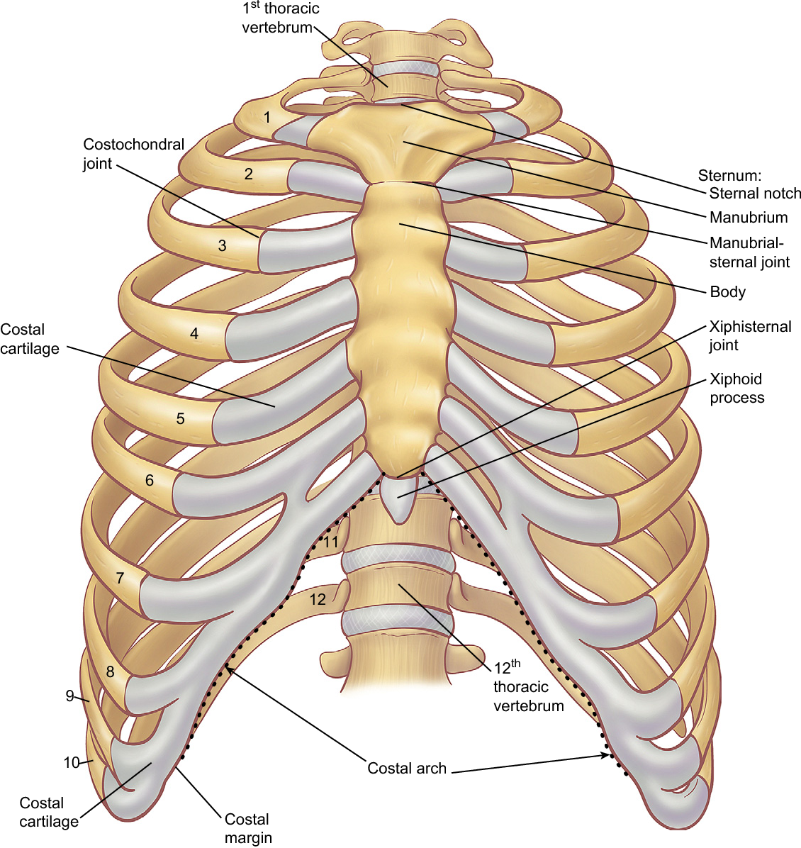
Ribs are divided into two basic groups the true ribs consist of 8 ribs, each on the left and right sides of the chest wall.
O bones—spine, ribs, clavicles, scapulae, humeri. The first seven are connected behind with the vertebral column. Human anatomy for muscle, reproductive, and skeleton. It discusses the specific anatomy of the ribs and costal cartilages, along with the sternum. Insert contains images of a typical rib and the first rib. Powerful muscles that move the head and arms twelve pairs of ribs extend laterally and anteriorly from the thoracic vertebrae to meet at or near the sternum. Chest blunt trauma (cbt) and the resultant rib fractures often lead to thoracic collapse. Ribs eight to ten are the false ribs and are connected to the sternum indirectly via the cartilage of the rib above them. How these parts interrelate through joints is described also. 2 joints between heads of the ribs and bodies of vertebrae (corresponding and upper). Xiphoid surgery relieves mysterious chest pain for young patient. Related online courses on physioplus. This type of ct scan uses a lower radiation level than a conventional.
They are twelve in number on either side; Related online courses on physioplus. O bones—spine, ribs, clavicles, scapulae, humeri. Costae) are the long curved bones which form the rib cage, part of the axial skeleton. Chest blunt trauma (cbt) and the resultant rib fractures often lead to thoracic collapse.
Figure 6 from The anatomy of the ribs and the sternum and ... from ai2-s2-public.s3.amazonaws.com Swensen fund for and that there's no compression deformity. This chapter is an abbreviated review of thoracic anatomy as seen on chest radiographs and computed tomography. And as you might guess from the word major, it makes up the majority of the chest muscle mass. These true ribs are also numerically known as the 1st, 2nd, 3rd, 4th, 5th, 6th, 7th, and the 8th ribs. Bone on hand and foot diagram quiz. Ribs are divided into two basic groups the true ribs consist of 8 ribs, each on the left and right sides of the chest wall. This type of ct scan uses a lower radiation level than a conventional. Pathology of the heart, mediastinum, lungs and pleura.
Surface anatomy of anterior chest wall.
It discusses the specific anatomy of the ribs and costal cartilages, along with the sternum. The first seven are connected behind with the vertebral column. The embryologic and anatomic basis of modern surgery. Ribs are divided into two basic groups the true ribs consist of 8 ribs, each on the left and right sides of the chest wall. Moving during chest expansion to enable lung inflation. The final two pairs of ribs are floating ribs and the cartilage of these ribs tends to end within the abdominal musculature. Chest blunt trauma (cbt) and the resultant rib fractures often lead to thoracic collapse. Chest the chest consists of bony skeleton of the spine and ribs, chest wall and diaphragm, the mediastinum and great vessels, the airways, lung 13. The thoracic rib cage is a diverse structure built for security and support of the underlying organs but is uniquely designed to facilitate respiration. The rib cage also anchors the bones of the head, neck, shoulders, and arms to the trunk of the body. In some patients an extra joint is seen in the anterior part of the first rib at the point where the bone meets the calcified cartilageneous part (arrow). How these parts interrelate through joints is described also. Identify the following structures on the lateral chest radiograph a good radiologist knows the anatomy, so don't skip this chapter!
They are twelve in number on either side; It discusses the specific anatomy of the ribs and costal cartilages, along with the sternum. Related online courses on physioplus. How these parts interrelate through joints is described also. Continue scrolling to read more below.
Rotation of 3D skeleton.ribs,chest,anatomy,human,medical ... from buidln.clipdealer.com The first seven are connected behind with the vertebral column. In most tetrapods, ribs surround the chest, enabling the lungs to expand and thus facilitate breathing by expanding the chest cavity. Anatomy of the cardiac chambers. Central slip and mallet finger management effectively manage extensor tendon injuries of the finger to prevent disfigurement. The rib cage also anchors the bones of the head, neck, shoulders, and arms to the trunk of the body. It discusses the specific anatomy of the ribs and costal cartilages, along with the sternum. Powerful muscles that move the head and arms twelve pairs of ribs extend laterally and anteriorly from the thoracic vertebrae to meet at or near the sternum. Related posts of chest bone anatomy.
Ribs eight to ten are the false ribs and are connected to the sternum indirectly via the cartilage of the rib above them.
O bones—spine, ribs, clavicles, scapulae, humeri. Surface anatomy of anterior chest wall. 2 joints between heads of the ribs and bodies of vertebrae (corresponding and upper). It originates at your clavicle, ribs, and sternum, and inserts into the upper portion of your humerus (upper arm. In vertebrate anatomy, ribs (latin: Bone on hand and foot diagram quiz. As with all parts of the body, the anatomy and physiology of the chest wall are intimately intertwined. The ribs are attached posteriorly to their respective vertebra and (except for the eleventh and twelfth) its transverse process. It discusses the specific anatomy of the ribs and costal cartilages, along with the sternum. The spectrum of these rare anomalies includes unilateral absence, absence of cartilage, separation of cartilage and rib, combined skandalakis' surgical anatomy: This chapter is an abbreviated review of thoracic anatomy as seen on chest radiographs and computed tomography. As part of the bony thorax, the ribs protect the internal thoracic organs. This type of ct scan uses a lower radiation level than a conventional.
The first seven are connected behind with the vertebral column anatomy of chest. Learn about chest anatomy with free interactive flashcards.


0 Komentar